Akshara G.
- Sep 18, 2025
- 3 min read
This summer, I had the incredible opportunity to be part of the Fred Hutch Pathways Explorers Program. It was an experience that opened the doors to the world of biomedical research and gave me the chance to try so many new things. Over the course of the program, I gained new lab skills, explored different research areas, and found a field of science that I’m truly passionate about.
One of the first things we learned was how to use some of the core techniques that scientists rely on every day. I ran my first gel electrophoresis, which separates DNA fragments by size, used PCR (Polymerase Chain Reaction) to amplify DNA, and even used CRISPR Cas9 to edit genetic material like a BRCA-1 tumor suppressor gene. These are tools that are essential to modern biology and getting the chance to actually do these experiments myself felt both challenging and exciting.

My gel from a gel electrophoresis experiment. The bands show fragments separated by charge and size using an electric current
We also received the opportunity to visit a variety of labs on campus, each with its own focus. Some of the most memorable stops were the C. elegans lab, the Fruit Fly Lab (which studies single cell wound healing), the Zebrafish Lab, the Genomics Core, and the Histopathology Core. Each one showed me a different way science can answer questions about health and disease, and I left with a better understanding of how broad the field of biomedical research really is.
However, the part of the program that stood out to me the most was the structural biology lecture by Dr. Stoddard. He explained how scientists use structural biology to see proteins at the atomic level and essentially created blueprints of how they work. This kind of research is critical for developing new medicines because it shows exactly how molecules fit together and interact. His talk made me realize how much I enjoyed thinking about science in this way, and it sparked a passion I hadn’t expected.
After the lecture, we visited Dr. Stoddard’s lab, where I got to see x-ray protein crystallography up close. This technique uses x-rays and protein crystals to build 3D models of proteins. It does this by firing x-rays at a protein crystal such that the machine is able to collect all of the diffraction patterns (which is why it’s important to have big crystals!) and put it through a mathematical equation to create a 3D model of the protein. Seeing how something so complex could be visualized in such detail was amazing, and it felt like physics and biology were coming together to solve the mysteries of proteins and life.

Protein crystals under the microscope, which are used in x-ray crystallography to determine the 3D shapes of proteins

The x-ray protein crystallography machine in Dr. Stoddard’s lab. It fires x-rays at protein crystals and collects diffraction patterns, which scientists analyze to reveal the protein’s structure
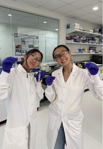
Beyond science, I also learned a lot about collaboration. Working with other students, sharing ideas, and working through experiments together reminded me that science is a team effort, and research depends not just on curiosity, but also on communication and teamwork.
Looking back, the Pathways Explorers Program gave me both practical skills and inspiration. From running my first gel to standing in a structural biology lab, every moment was meaningful. Most importantly, it helped me discover my passion for structural biology, and I will use that finding for years to come!


Comments